(Showing 1 - 21 products of 21 products)
Categories
Sub Categories
Brands
Suppliers
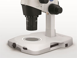
Microscope Stereo 0.8-4x zoom sz51-set olympus
MOQ: 1
Greenough Optical System Ray Diagram SZ51/61 have zoom ratios of 5:1 and 6.7:1 (zoom range 0.8x-4x and 0.67x-4.5x) respectively and are affordably priced yet off the highest resolving power in their price range and are available with either 45 or 60 degree inclination angles for observation. Utilizing the Greenough optical design, all zoom bodies incorporate the latest aberration and distortion-free optics that set a new standard in optical quality while maintaining superior operability.The V-shape optical path ensures a slim zoom body - ideal for integration into other equipment or stand alone use.3 Dimensional Viewing The optimum inward angle allows just the right combination of high level flatness and depth of focus for 3D viewing. Even a specimen with significant depth can be brought into focus from top to bottom for faster inspectionObservation Images with High Color Fidelity Superior optical coatings render true color images with fine detail.ESD Measures for Protecting Devices from Static ElectricityThe main unit and major accessories can quickly eliminate static electricity with the use of antistatic materials and coatings. This prevents a specimen under observation from electrostatic damage.
OLYMPUS SZ61 MICROSCOPE SPECIFICATIONS: | |
---|---|
Head: | Binocular 45 degree |
Zoom Range: | 0.67x - 4.5x |
Eyepieces: | 10x/22mm FOV |
Overall Mag: | 6.7x - 45x |
Working Distance: | 110mm |
Stand: |
Plain Stand LED Transmitted & Reflected Boom Stand Ball Bearing Boom Stand (Other Options Available Please Contact Us - All Generic Stands) |
Wholesaler/Dealer/Distributor
Kupondole, Lalitpur
Olympus Microscope IX73 with Narishige ON4 Micromanipulator
MOQ: 1
The Olympus IX73 inverted microscope has been integrated into our IXplore systems. IXplore Systems are designed to provide solutions-based packages that suit your research application needs.
Expandable to Meet Growing Research Needs
The semi-motorized IX73 is designed to satisfy a variety of research needs. With two deck options and additional modules for expanded functionality, the IX73 is perfectly suited for a changing research environment.
IX73: Two-deck System
The modular IX73 two-deck system can be combined with coded or motorized units for maximum expandability.
IX73: One-deck System
A microscope designed with an emphasis on efficiency. Ideal for documentation, routine testing and other tasks.
Reliable, Clear and Bright High-Resolution Images
Olympus UIS2 infinity-corrected optics ensure high optical transmittance with a broad range of objectives. UIS2 optics feature wide chromatic correction and enable high resolution, high S/N primary images regardless of the observation method. The wide field of view and Fly-Eye lens system provide uniform fluorescence images and enable the use of sCMOS cameras with large sensors.
Excellent Image Quality
Apochromatic Objectives Enable High-Resolution Phase Contrast and Fluorescence Observation
Phase contrast apochromatic objectives (UPLSAPO100XOPH, PLAPON60XOPH) enable high-precision imaging free from image shift, even during simultaneous phase contrast and fluorescence observation. This eliminates the need to change objectives when switching observation methods.
Wholesaler/Dealer/Distributor
Kupondole, Lalitpur
Olympus CX23 Upright Microscope
MOQ: 1
User Friendly
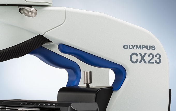
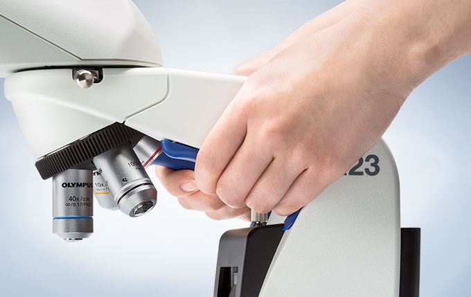
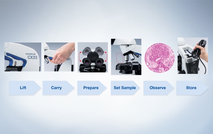
Ergonomically designed, the CX23’s molded grips add comfort and safety when retrieving the microscope from high places, such as shelves. Additionally, their blue color clearly indicates where to hold the microscope.
The CX23 is one of the lightest weight microscope in its class, with a total weight of approx. 5.9 kg(13.01 lbs).
The CX23 features a carrying arm angled to keep the wrists in a comfortable, neutral position.
The eyepieces can be fixed in place to help prevent damage or loss during transportation.
A rotational binocular tube can be locked for secure positioning.
Interpupillary adjustments, ranging from 48 to 75 mm, allowsindividual users to set to their needs. Likewise, eyepoint adjustments accommodate the user’s height and provides greater comfort.
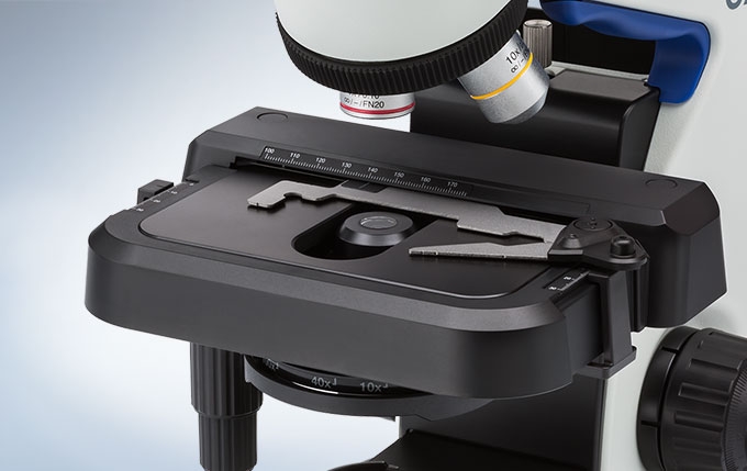
A rackless stage and stage cover give safety and stable longtime operation.
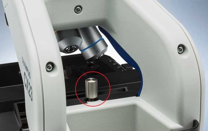
The focus lock prevents objectives and specimen from any damage.
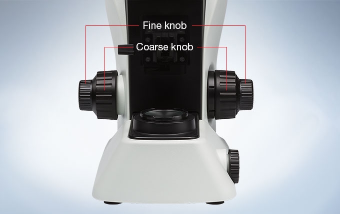
Bring specimens quickly into focus by using the coaxial knobs for fine or coarse adjustment. Durably built for movement and precise control, whether accessed from the left or right side.
Left and right diopter adjustment enable optimal focus for each eye.
Easily set and exchange specimens or add oil for the immersion objective with the CX23's inward-facing revolving nosepiece design. Allowing for more space above the stage, this design also protects specimens from objectives with a long working distance.
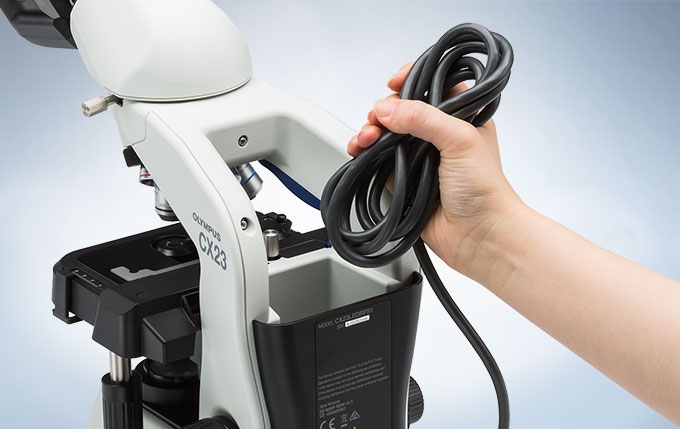
A storage compartment at the back of the CX23 makes it easy to stow the power cable after use.
Protect your investment further with an optional custom designed wooden case.
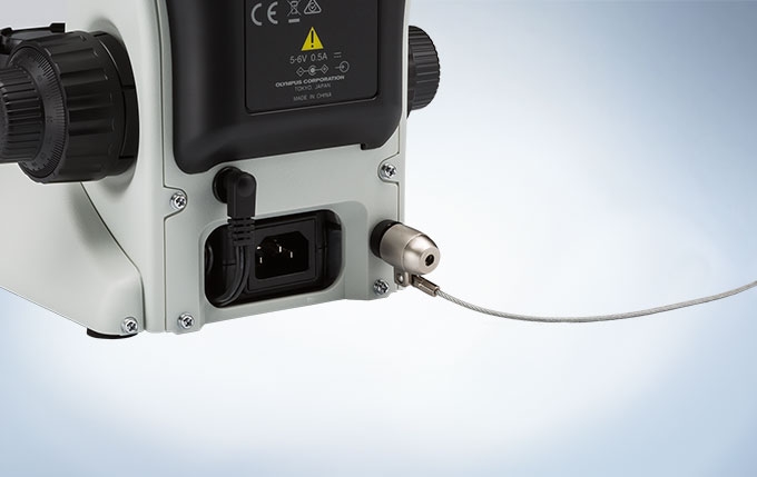
For added reassurance in situations when the CX23 needs to be left unattended, a built-in security slot allows attachment of an antitheft cable.
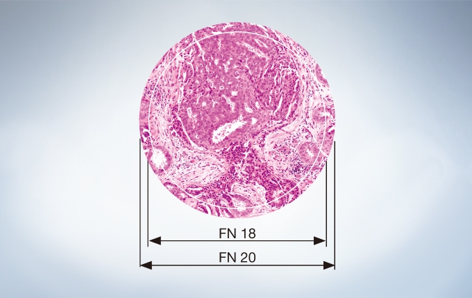
The CX23 allows users to see more with a greater field number than standard microscopes in this class.
The CX23 features plan achromat objectives that preserve outstanding image flatness throughout the field of view.
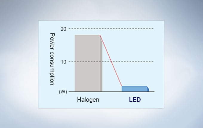
The LED light source provides long life time of 20,000 hours, and offers low power consumption. Also, its reduced blue color preserves vivid colors on HE staining.
Damage when excessive force is applied to the coarse focus or stage knob is prevented with a built-in torque release function.
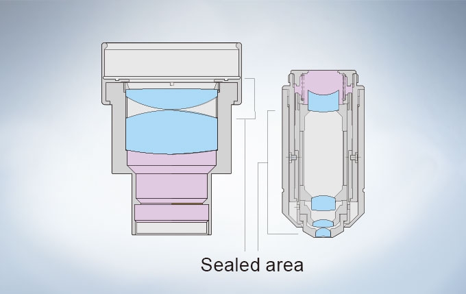
Many microscopes are vulnerable to mold and other fungi when used in hot, humid environments. The objectives, eyepieces, and observation tube of the CX23 have all undergone anti-fungus treatment, enabling optimal viewing and greater durability.
Observation Method | Brightfield | ? | |
Darkfield | ? | ||
Illuminator | Transmitted Koehler Illuminator |
LED Lamp | ? |
Focus | Focusing Mechanism | Stages Focus | ? |
Coarse Handle Stroke | 15mm | ||
Features | Coarse adjustment limit stopper Torque adjustment for coarse adjustment knob |
||
Revolving Nosepiece |
Manual | Standard Types | Built-in 4 position |
Stage | Mechanical | Mechanical Stages with Right-Hand Control | Built-in X: 76 mm, Y: 30 mm |
Condenser | Manual | Abbe Conenser | NA1.25/ W.D.- (4 X - 100 X) (Built-in) |
Observation Tubes | Standard (FN20) | Binocular | ? |
Trinocular | ? | ||
Tube Inclination Angle | 30° | ||
Trinocular Tube Light Path Selection (Camera : Observation) | 0 % : 100 %, 100 % : 0 % | ||
Interpupillary Distance Adjustment | 48 - 75 mm | ||
Dimensions | 198 (W) x 258 (D) x 384 (H) mm | ||
Weight | Approx. 5.9 kg |
Wholesaler/Dealer/Distributor
Kupondole, Lalitpur
Olympus BX53 Upright Microscope
MOQ: 1
Scalability is the keyword for this microscope. It is designed as a modular system allowing for the addition of many peripherals and applications to meet varying job requirements.
Most of the accessories for other Olympus microscopes are available for the BX53 including:
Olympus microscopes are noted for versatility and the scalability of this unit will allow for accessory modules to be added to meet currently unanticipated needs.
The lack of full computer integration in a unit designed for high end professional use is disappointing.
Summary:
Main Components:
Observation Tube:
Objective Lenses:
Fluorescence Illuminator.
Fluorescence Filter Cubes:
These three filter cubes are like-new:
U-FGNA
U-FBN
U-FUN
We can provide customized filter sets as per customer’s requirements. We are a dealer for Chroma. Chroma supplies Olympus with their dichroic filters.
Misc Notes:
History of this Instrument:
Options:
SPECIFICATION | |
---|---|
OPTICAL SYSTEM | UIS2 optical system |
FOCUS | Coaxial coarse and fine focus with stage up and down mechanism Focus stroke 25 mm Coarse stroke 15 mm/rotation Fine stroke 100 μm/rotation Graduation on fine focus 1 μm Prefocusing limit stopper and torque adjustment on coarse focus |
ILLUMINATOR | Built-in Koehler illumination for transmitted light Light intensity LED indicator, Built-in filters (LBD-IF, ND6, ND25) 12 V, 100 W halogen bulb (pre-centered) |
REVOLVING NOSEPIECE | Interchangeable reversed quintuple/sextuple/septuple nosepiece Encoding optional with septuble nosepiece Motorized septuple revolving nosepiece |
OBSERVATION TUBE | Widefield tilting, telescopic and lifting binocular, inclined -3°-27° Widefield tilting trinocular, inclined 5°–35° Widefield trinocular, inclined 30° Widefield erect image trinocular, inclined 30° Widefield tilting binocular, inclined 5°–35° Widefield ergo binocular, inclined 0°–25° Widefield binocular, inclined 30° Super widefield trinocular, inclined 24° Super widefield erect image trinocular, inclined 24° |
STAGE | Ceramic-coated coaxial stage with left or right hand low drive control, with rotating mechanism and torque adjustment mechanism, optional ergo grips available (Non stick coated grooved coaxial, plain, rotatable stages are also available) |
CONDENSER | Swing out achromatic condenser (N.A. 0.9), for 1.25x–100x (swing-out: 1.25x–4x) Achromatic aplanatic condenser (N.A. 1.4), for 10x–100x Phase contrast, darkfield condenser (N.A. 1.1), [phase contrast: for 10x–100x, darkfield: for 10x–100x (up to N.A. 0.80)] Universal condenser (N.A. 0.9), for 1.25x–100x [swing-out: 1.25x–4x,with oil top lens:(N.A. 1.4)] Low magnification condenser (N.A. 0.75), for 2x–100x (Dry) Ultra low magnification condenser (N.A. 0.16), for 1.25x–4x Darkfield dry condenser (N.A. 0.8–0.92), for 10x–100x Darkfield oil condenser (N.A. 1.20–1.40), for 10x–100x Motorized universal condenser (N.A. 0.9, motorized 8-position turret, Aperture stop, polarizing filter in/out mechanism and top lens swing out mechanism), for 1.25x–100x [swing-out 1.25x-4x, with oil top lens: (N.A. 1.4)] |
FLUORESCENCE ILLUMINATOR | Manual reflected fluorescence, 8-position mirror turret unit, encoded with tool-free exchange of filter cubes Motorized reflected fluorescence, 8-position mirror turret unit, encoded with tool-free exchange of filter cubes |
FLUORESCENCE LIGHT SOURCE | 100 W Hg apo lamp housing and transformer 100 W Hg lamp housing and transformer 75 W Xe lamp housing and transformer Fibre coupled metal-halide lightsources Fibre coupled LED lightsource |
CONTROLLER | Optional: Control box for semi-motorised setups |
OPERATING ENVIRONMENT | Indoor use Ambient temperature: 5 ºC to 40 ºC (41 º0 to 104 º0) Maximum relative humidity: 80% for temperatures up to 31º C (88 º0), decreasing linearly through 70% at 34 ºC (93 º0), 60% at 37 ºC (99 º0), to 50% relative humidity at 40 ºC (104 º0) Supply voltage fluctuations: Not to exceed ±10% of the normal voltage |
Wholesaler/Dealer/Distributor
Kupondole, Lalitpur
Microscopic Stage Micrometer erma japan
MOQ: 1
BRAND NAME | Erma |
---|---|
EAN | 0008725953663 |
MODEL NUMBER | Stage Micrometer |
NUMBER OF ITEMS | 1 |
PART NUMBER | Objective micrometer |
UPC | 008725953663 |
Wholesaler/Dealer/Distributor
Kupondole, Lalitpur
Microscopic Reflector 50m
MOQ: 1
Structural coloration is common in dynamic visual displays by terrestrial and marine animals [1–5]. The spectral environment in which they live influences communication methods that use coloration [1,6–11]. In the ocean's euphotic zone, the ‘disco’ or ‘electric’ file clam Ctenoides ales (family Limidae) is found inside small crevices at depths of approximately 3–50 m. At these depths, the majority of wavelengths available for visual displays are in the blue-green range (400–500 nm) [12]. This is true even at shallow depths where long wavelengths have not yet attenuated (less than 15 m), as the crevices in which C. ales are found are dominated by horizontal light composed of short wavelengths (electronic supplementary material, file S1).
The flashing display on the mantle lip of C. ales has been mischaracterized as bioluminescence [13,14], although it is actually mediated by light scattered from photonic nanostructures [15]. Structures of this sort typically use materials that have high refractive indices relative to the substrate, such as collagen, chitin, keratin and guanine [16]. Silica also has a high refractive index (n = 1.43 at 589 nm) [16] but has only rarely been used as a biophotonic structure, such as in diatoms [17–19] and the weevil Pachyrhynchus argus [20]. Photonic nanostructures of any substance however can enhance reflectance, such as the ultrathin, aperiodic filaments in scales of Cyphochilus spp. beetles [21] and the bead-studded scales in the wings of certain pierid butterflies [22].
Within bivalves, studies of light manipulation are limited to the bioluminescence of the marine clams Pholas dactylus and Gastrochaena sp. [23], and the iridophores of the giant clam Tridacna, which are thought to scatter light towards symbiotic zooxanthallae [24]. Ctenoides ales, however, is the only known bivalve with a behaviourally mediated photic display. The fundamental characteristics of this display are described here to determine its potential as a signal. Ongoing studies of the function of the display are being conducted in the context of habitat-specific sensory ecology. In order to provide a preliminary comparative framework, we collected data from the morphologically and ecologically similar congener Ctenoides scaber, which does not flash.
Wholesaler/Dealer/Distributor
Kupondole, Lalitpur
Microscopic Ocular Micrometer 0.1 mm bestscope
MOQ: 1
Wholesaler/Dealer/Distributor
Kupondole, Lalitpur
Microscopic Eyepiece 10x
MOQ: 1
Eyepieces work in combination with microscope objectives to further magnify the intermediate image so that specimen details can be observed. Oculars is an alternative name for eyepieces that has been widely used in the literature, but to maintain consistency during this discussion we will refer to all oculars as eyepieces.
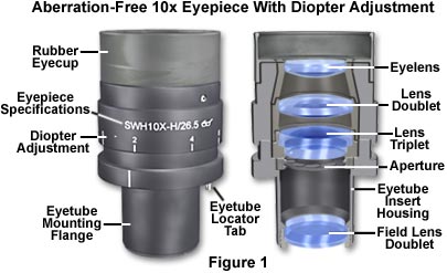
Best results in microscopy require that objectives be used in combination with eyepieces that are appropriate to the correction and type of objective. The basic anatomy of a typical modern eyepiece is illustrated in Figure 1. Inscriptions on the side of the eyepiece describe its particular characteristics and function.
The eyepieces illustrated in Figure 1 are inscribed with UW, which is an abbreviation for the Ultra Wide viewfield. Often eyepieces will also have an H designation, depending upon the manufacturer, to indicate a high-eyepoint focal point that allows microscopists to wear glasses while viewing samples. Other inscriptions often found on eyepieces include WF for Wide-Field; UWF for Ultra Wide-Field; SW and SWF for Super Wide-Field; HE for High Eyepoint; and CF for eyepieces intended for use with CF corrected objectives. Compensating eyepieces are often inscribed with K, C, or comp as well as the magnification. Eyepieces used with flat-field objectives are sometimes labeled Plan-Comp. The eyepiece magnification of the eyepieces in Figure 1 is 10x (indicated on the housing), and the inscription A/24 indicates the field number is 24, which refers to the diameter (in millimeters) of the fixed diaphragm in the eyepiece. These eyepieces also have a focus adjustment and a thumbscrew that allows their position to be fixed. Manufactures now often produce eyepieces having rubber eye-cups that serve both to position the eyes the proper distance from the front lens, and to block room light from reflecting off the lens surface and interfering with the view.
There are two major types of eyepieces that are grouped according to lens and diaphragm arrangement: the negative eyepieces with an internal diaphragm and positive eyepieces that have a diaphragm below the lenses of the eyepiece. Negative eyepieces have two lenses: the upper lens, which is closest to the observer's eye, is called the eye-lens and the lower lens (beneath the diaphragm) is often termed the field lens. In their simplest form, both lenses are plano-convex, with convex sides "facing" the specimen. Approximately mid-way between these lenses there is a fixed circular opening or internal diaphragm which, by its size, defines the circular field of view that is observed in looking into the microscope.
The simplest negative eyepiece design, often termed the Huygenian eye-piece (illustrated in Figure 2), is found on most teaching and laboratory microscopes fitted with achromatic objectives. Although the Huygenian eye and field lenses are not well corrected, their aberrations tend to cancel each other out. More highly corrected negative eyepieces have two or three lens elements cemented and combined together to make the eye lens. If an unknown eyepiece carries only the magnification inscribed on the housing, it is most likely to be a Huygenian eyepiece, best suited for use with achromatic objectives of 5x-40x magnification.
The other main kind of eyepiece is the positive eyepiece with a diaphragm below its lenses, commonly known as the Ramsden eyepiece, as illustrated in Figure 2 (on the left). This eyepiece has an eye lens and field lens that are also plano-convex, but the field lens is mounted with the curved surface facing towards the eye lens. The front focal plane of this eyepiece lies just below the field lens, at the level of the eyepiece diaphragm, making this eyepiece readily adaptable for mounting reticles. To provide better correction, the two lenses of the Ramsden eyepiece may be cemented together.
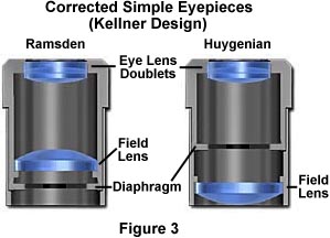
A modified version of the Ramsden eyepiece is known as the Kellner eyepiece, as illustrated on the left in Figure 3. These improved eyepieces contain a doublet of eye-lens elements cemented together and feature a higher eyepoint than either the Ramsden or Huygenian eyepiece as well as a much larger field of view. A modified version of the simple Huygenian eyepiece is also illustrated in Figure 3, on the right. While these modified eyepieces perform better than their simple one-lens counterparts, they are still only useful with low-power achromat objectives.
Simple eyepieces such as the Huygenian and Ramsden and their achromatized counterparts will not correct for residual chromatic difference of magnification in the intermediate image, especially when used in combination with high magnification achromatic objectives as well as any fluorite or apochromatic objectives. To remedy this, manufacturers produce compensating eyepieces that introduce an equal, but opposite, chromatic error in the lens elements. Compensating eyepieces may be either of the positive or negative type, and must be used at all magnifications with fluorite, apochromatic and all variations of plan objectives (they can also be used to advantage with achromatic objectives of 40x and higher). In recent years, modern microscope objectives have their correction for chromatic difference of magnification either built into the objectives themselves (Olympus and Nikon) or corrected in the tube lens (Leica and Zeiss).
Compensating eyepieces play a crucial role in helping to eliminate residual chromatic aberrations inherent in the design of highly corrected objectives. Hence, it is preferable that the microscopist uses the compensating eyepieces designed by a particular manufacturer to accompany that manufacturer's higher-corrected objectives. Use of an incorrect eyepiece with an apochromatic objective designed for a finite (160 or 170 millimeter) tube length application results in dramatically increased contrast with red fringes on the outer diameters and blue fringes on the inner diameters of specimen detail. Additional problems arise from a limited flatness of the viewfield in simple eyepieces, even those corrected with eye-lens doublets.
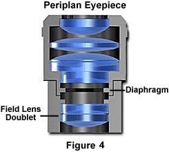
More advanced eyepiece designs resulted in the Periplan eyepiece that is illustrated in Figure 4 above. This eyepiece contains seven lens elements that are cemented into a single doublet, a single triplet, and two individual lenses. Design improvements in periplan eyepieces lead to better correction for residual lateral chromatic aberration, increased flatness of field, and a general overall better performance when used with higher power objectives.
Modern microscopes feature vastly improved plan-corrected objectives in which the primary image has much less curvature of field than older objectives. In addition, most microscopes now feature much wider body tubes that have greatly increased the size of intermediate images. To address these new features, manufacturers now produce wide-eyefield eyepieces (illustrated in Figure 1) that increase the viewable area of the specimen by as much as 40 percent. Because the strategies of eyepiece-objective correction techniques vary from manufacturer to manufacturer, it is very important (as stated above) to use only eyepieces recommended by a specific manufacturer for use with their objectives.
Our recommendation is to carefully choose the objective first, then purchase an eyepiece that is designed to work in conjunction with the objective. When choosing eyepieces, it is relatively easy to differentiate between simple and more highly compensated eyepieces. Simple eyepieces such as the Ramsden and Huygenian (and their more highly corrected counterparts) will appear to have a blue ring around the edge of the eyepiece diaphragm when viewed through the microscope or held up to a light source. In contrast, more highly corrected compensating eyepieces with have a yellow-red-orange ring around the diaphragm under the same circumstances.
Wholesaler/Dealer/Distributor
Kupondole, Lalitpur
Microscopic Camera Eyepiece with Micromter for stero microscpe
MOQ: 1
Model:MDE4-500BC
Sensor & Size(mm):5.0M/SC5033(C) 1/2.7"(5.18x3.89)
Pixel(μm):2.0x2.0
FPS/Resolution:15@2560x1920,20@2048x1536,20@1600x1200,30@800x600
Binning:1x1,2x2
Exposure:Auto
Spectral Range:380-650nm
White Balance:Auto White Balance
Packaging Details:Strong Carton with Polyfoam Protection
Port:Beijing
Lead Time:Within 1-2 weeks after receiving payment
MDE4-500BC(With SC5033(C) Sensor) is an economic version with simple and compact structure USB3.0 CMOS eyepiece camera. USB3.0 is used as the data transfer interface.
Microscope eyepiece camera with 23.2 diameter and compact size;
The MDE4 comes with high-speed USB3.0 interface and high frame rate video display keep the screen smooth without interruption; Also the MDE4 comes with advanced video & image processing application ImageView.
The MDE4 can be widely used to transfer the mono or binocular student microscopes to digital microscope.
With 23.2 to 30mm or 23.2 to 30.75 convert ring, the MDE4 camera can also change the stereo microscope to digital stereo microscope.
The basic characteristic of MDE4 cameras are as follows:
Order Code |
Sensor & Size |
Pixel(μm) |
G Responsivity Dynamic range SNRmax |
FPS/Resolution |
Binning |
Exposure |
MDE4-500BC |
5.0M/SC5033(C) 1/2.7″(5.18×3.89) |
2.0×2.0 |
2 V/lux-sec 64dB 35dB |
15@2560×1920 30@800×600 |
1×1,2×2 |
Auto |
C: Color; M: Monochrome;
Other Specification for MDE4 Camera |
|
Spectral Range |
380-650nm (with IR-cut Filter) |
White Balance |
Auto White Balance |
Color Technique |
N/A |
Capture/Control API |
Native C/C++, C#/VB.NET, DirectShow, Twain and Labview |
Recording System |
Still Picture and Movie |
Cooling System* |
Natural |
Operating Environment |
|
Operating Temperature (in Centigrade) |
-10~ 50 |
Storage Temperature (in Centigrade) |
-20~ 60 |
Operating Humidity |
30~80%RH |
Storage Humidity |
10~60%RH |
Power Supply |
DC 5V over PC USB3.0 Port |
Software Environment |
|
Operating System |
Microsoft® Windows® XP / Vista / 7 / 8 /10 (32 & 64 bit) OSx(Mac OS X) Linux |
PC Requirements |
CPU: Equal to Intel Core2 2.8GHz or Higher |
Memory:2GB or More |
|
USB Port:USB3.0 High-speed Port |
|
Display:17” or Larger |
|
CD-ROM |
The MDE4 body, made from aluminum alloy blackening, ocular housing: Dia.32 X 56mm ensures a heavy duty, workhorse solution. The camera is designed with a high quality IR-CUT filter to filter the infrared light and protect the camera sensor. No moving parts included. This design ensures a rugged, robust solution with an increased lifespan when compared to other industrial camera solutions.
Standard Camera Packing List |
||
A |
Carton L:52cm W:32cm H:33cm (20pcs, 12~17Kg/ carton), not shown in the photo |
|
B |
Gift box L:15cm W:15cm H:10cm (0.25~0.35Kg/ box) |
|
C |
MDE4 series camera |
|
D |
High-Speed USB3.0 USB315-ATA | USB 3.0 A Male to A Male Cable,1.5m |
|
E |
CD (Driver & utilities software, Ø8cm) |
|
Optional Accessory |
||
0 |
C-Mount Adapter Housing:108027(HS502) |
|
G |
108015(Dia.23.2mm to 30.0mm Ring)/Adapter rings for 30mm eyepiece tube |
|
H |
108016(Dia.23.2mm to 30.5mm Ring)/ Adapter rings for 30.5mm eyepiece tube |
|
I |
108017(Dia.23.2mm to 31.75mm Ring)/ Adapter rings for 31.75mm eyepiece tube |
|
J |
Calibration kit |
106011/TS-M1(X=0.01mm/100Div.)? |
Wholesaler/Dealer/Distributor
Kupondole, Lalitpur
Microscopic Camera Eyepiece 2mp bestscope
MOQ: 1
Model:MDE4-500BC
Sensor & Size(mm):5.0M/SC5033(C) 1/2.7"(5.18x3.89)
Pixel(μm):2.0x2.0
FPS/Resolution:15@2560x1920,20@2048x1536,20@1600x1200,30@800x600
Binning:1x1,2x2
Exposure:Auto
Spectral Range:380-650nm
White Balance:Auto White Balance
Packaging Details:Strong Carton with Polyfoam Protection
Port:Beijing
Lead Time:Within 1-2 weeks after receiving payment
MDE4-500BC(With SC5033(C) Sensor) is an economic version with simple and compact structure USB3.0 CMOS eyepiece camera. USB3.0 is used as the data transfer interface.
Microscope eyepiece camera with 23.2 diameter and compact size;
The MDE4 comes with high-speed USB3.0 interface and high frame rate video display keep the screen smooth without interruption; Also the MDE4 comes with advanced video & image processing application ImageView.
The MDE4 can be widely used to transfer the mono or binocular student microscopes to digital microscope.
With 23.2 to 30mm or 23.2 to 30.75 convert ring, the MDE4 camera can also change the stereo microscope to digital stereo microscope.
The basic characteristic of MDE4 cameras are as follows:
Order Code |
Sensor & Size |
Pixel(μm) |
G Responsivity Dynamic range SNRmax |
FPS/Resolution |
Binning |
Exposure |
MDE4-500BC |
5.0M/SC5033(C) 1/2.7″(5.18×3.89) |
2.0×2.0 |
2 V/lux-sec 64dB 35dB |
15@2560×1920 30@800×600 |
1×1,2×2 |
Auto |
C: Color; M: Monochrome;
Other Specification for MDE4 Camera |
|
Spectral Range |
380-650nm (with IR-cut Filter) |
White Balance |
Auto White Balance |
Color Technique |
N/A |
Capture/Control API |
Native C/C++, C#/VB.NET, DirectShow, Twain and Labview |
Recording System |
Still Picture and Movie |
Cooling System* |
Natural |
Operating Environment |
|
Operating Temperature (in Centigrade) |
-10~ 50 |
Storage Temperature (in Centigrade) |
-20~ 60 |
Operating Humidity |
30~80%RH |
Storage Humidity |
10~60%RH |
Power Supply |
DC 5V over PC USB3.0 Port |
Software Environment |
|
Operating System |
Microsoft® Windows® XP / Vista / 7 / 8 /10 (32 & 64 bit) OSx(Mac OS X) Linux |
PC Requirements |
CPU: Equal to Intel Core2 2.8GHz or Higher |
Memory:2GB or More |
|
USB Port:USB3.0 High-speed Port |
|
Display:17” or Larger |
|
CD-ROM |
The MDE4 body, made from aluminum alloy blackening, ocular housing: Dia.32 X 56mm ensures a heavy duty, workhorse solution. The camera is designed with a high quality IR-CUT filter to filter the infrared light and protect the camera sensor. No moving parts included. This design ensures a rugged, robust solution with an increased lifespan when compared to other industrial camera solutions.
Standard Camera Packing List |
||
A |
Carton L:52cm W:32cm H:33cm (20pcs, 12~17Kg/ carton), not shown in the photo |
|
B |
Gift box L:15cm W:15cm H:10cm (0.25~0.35Kg/ box) |
|
C |
MDE4 series camera |
|
D |
High-Speed USB3.0 USB315-ATA | USB 3.0 A Male to A Male Cable,1.5m |
|
E |
CD (Driver & utilities software, Ø8cm) |
|
Optional Accessory |
||
0 |
C-Mount Adapter Housing:108027(HS502) |
|
G |
108015(Dia.23.2mm to 30.0mm Ring)/Adapter rings for 30mm eyepiece tube |
|
H |
108016(Dia.23.2mm to 30.5mm Ring)/ Adapter rings for 30.5mm eyepiece tube |
|
I |
108017(Dia.23.2mm to 31.75mm Ring)/ Adapter rings for 31.75mm eyepiece tube |
|
J |
Calibration kit |
106011/TS-M1(X=0.01mm/100Div.)? |
Wholesaler/Dealer/Distributor
Kupondole, Lalitpur
Microscopic Camera Digital hdmi/wifi/gige 8.0mp with adapter bwhc-4k8mpb bestscope
MOQ: 1
Introduction
BWHC-4K8MPA 4K UHD WIFI HDMI digital camera has adopted high sensitivity 1/1.8 inch 8.3MP Sony CMOS image sensor, pixel size 2.0um. The camera has high dynamic range, high sensitivity and excellent thermal noise suppression features. The camera outputs 4K resolution of 3840 x2160 pixels, the resolution is 4 times as 1080P cameras, maximum frame rate is 60fps. The Camera can be connected to 4K UHD Screen via HDMI interface, it also can be connected to PC via USB3.0, WIFI or GigE and controlled with professional software. The captured images will be saved to SD card or USB flash drive, the camera can ensure every detail is not to be missed.
BWHC-4K8MPA can be controlled by a mouse, the operation is simple and quick. This camera has many measurement functions, like point, line, circle, rectangle, annulus, polygon, polyline, angle, double circle, distance from point to line, concentric circles and so on.
Application
BWHC-4K8MPA 4K UHD WIFI HDMI digital camera can be widely used in video conferencing, remote medical diagnosis, microscopy images, industrial inspection, video projectors, security monitoring field. With the high image quality and easy to operate features, it will be your best assistant for following applications:
BWHC-4K8MPA Datasheet
Order Code | Sensor Type (mm) | Pixel Size (μm) | G-sensitivity | FPS/Resolution | Binning | Exposure Time |
BWHC-4K8MPA | Sony IMX334(C) 1/1.8"(7.68x4.32) |
2.0x2.0 | 505mv with 1/30s 0.15mv with 1/30s |
3840*2160/60fps | 1x1 | 0.04ms~1000ms |
C:color; M:Monochrome;
BWHC-4K8MPA Interface Description
HDMI | For 4k/1080P video image output. Compliant with HDMI2.0. | |
USB Mouse | For connecting USB Mouse. Used for controlling the HDMI camera with XCamView | |
USB 3.0 | USB flash drive | |
Connecting 5G Wi-Fi adapter for wireless video image transmission (will be added in second version) | ||
USB video image output (will be added in second version) | ||
GE | Gigabit Ethernet port connecting router/switch for network transmission of video images | |
SD | SD-card slot. Compliant with SDIO3.0. | |
DC12V | 12V Power in | |
ON/OFF | Power On/ Off switch | |
LED | Power Indicator |
BWHC-4K8MPA Function Description
1. Video Image Output
Video Image Output | HDMI interface 4k/60HZ or 1080P/60HZ output |
Gigabit Ethernet port 4k/60FPS H264 encoded video image output. Support real-time adjustment of encoding bandwidth. Support network unicast/multicast configuration. Support DHCP acquisition and manual network configuration. Convenient for different network requirements. | |
Connecting USB3.0 5G Wi-Fi adapter to realize AP/ STA modes (will be added in a second version) | |
Connecting to computer by using USB3.0 cable to realize real-time MJPEG image output (will be added in a second version) |
2. Video Record and Image Capture
Video Record and Image Capture | Video Record: MP4 encapsulated H264 encoded 8MP (3840*2160 video, storage frame rate at 50~60FPS (depending on speed class of the SD Card and HDMI output resolution) |
Image Capture: 8.0MP(3840*2160) JPEG format |
Test Data:
Code | Test Condition | Test Result |
1 | 1.sensor 60FPS output (with exposure time limited within 16ms) 2.Using SanDisk SD card with speed class of CLASS 10 3.HDMI interface connecting to 4K displayer |
Storage frame rate: 50FPS |
2 | 1.sensor 60FPS output (with exposure time limited within 16ms) 2.Using SanDisk SD card with speed class of CLASS 10 3.HDMI interface connecting to 1080P displayer |
Storage frame rate: 60FPS |
3. ISP Function
ISP Function | Exposure Time/ Gain: Manual/ Auto Exposure. (Four auto-exposure strategies will be added in later version) |
White Balance: Manual/ Auto/ ROI White Balance | |
Sharpness | |
3D De-noise | |
Saturation | |
Contrast | |
Brightness | |
Gamma Curve | |
50/60HZ |
4. Image Assist Function
Image Assist Function | Zoom: 10 times digital zoom |
Mirror/Flip | |
Freeze | |
Cross line | |
PIP (Picture in Picture) | |
Compare: Comparison of real-time video and stored captured images | |
Image Browse | |
Video Playback | |
Measurement |
5. Others
Others | Built-in RTC clock providing accurate system time |
Restore Factory Setting function | |
Multiple language support (English, Simplified Chinese, Traditional Chinese, Korean, Thai) | |
Formats supported for SD Card/ USB flash drive: FAT32/EXFAT/NTFS |
Sample Images
Wholesaler/Dealer/Distributor
Kupondole, Lalitpur
Microscopic Camera Digital Hdmi/Wifi/Gige 16.0mp with adapter bwhc-4k8mpb bestscope
MOQ: 1
Model:BWHC-4K8MPA
Sensor & Size(mm):1/1.8"
Pixel(μm):2.0x2.0
G Sensitivity Dark Signal:505mv with 1/30s
FPS/Resolution:60@3840*2160(HDMI)
Binning:1x1
Exposure:0.04ms~1000ms
USB Port:USB3.0
Image Capture:8MP
Packaging Details:Strong Carton with Polyfoam Protection
Port:Beijing
Lead Time:Within 1-2 weeks after receiving payment
BWHC-4K8MPA 4K UHD HDMI/GigE/WiFi Multi-outputs Digital Camera(With Sony IMX334(C) Sensor) is the next-generation live-view imaging-system with 4K resolution at 60/30 FPS.
BWHC-4K series comes with Sony Exmor CMOS sensor with high sensitivity, low dark current and no smear achieved through the adoption of R, G and B primary color mosaic filters.
The cameras use a standard C-mount interface for maximum compatibility with various microscopy-systems. It can be used as a stand-alone recorder when used with an HDMI monitor or television, or live-streamed to a PC via Gigabit Ethernet (GE) for image-capture and video-recording.
Hardware 3D denoising, sharpness and local tone mapping control functions greatly improve the image and video quality.
The included Windows software offers image-development and measurement tools, as well as advanced compositing features such as image-stitching and extended-depth-of-focus. With the ability to calibrate scales at multiple magnifications, the software can be used for multi-level inspection. For Mac and Linux, there is a lite version of the software which can capture video and still images, and includes limited processing features.
The BWHC-4K series cameras are intended to be used for the acquisition of digital images from the stereo microscope, biological microscope or online interactive teaching.
The basic characteristics are listed as below:
1.Sony Exmor CMOS sensor;
2.4K HDMI/GE/WLAN multiple video outputs;
3.4K/1080P auto switching according to the display resolution;
4.SD card/USB flash disk for the captured image and video storage;
5.Embedded XCAMView for the control of the camera with mouse, no shaking;
6.With strong ISP and other related processing functions;
1.Scientific research, digital teaching (teaching, demonstration and academic discussing);
2.Digital laboratory, medical research;
3.Machine vision (PCB detection, IC quality control);
4.Medical treatment (pathological observation);
5.Food (microbial colony observation and counting);
6.Aviation and military.
Order Code |
Sensor & Size(mm) |
Pixel(μm) |
G Sensitivity Dark Signal |
FPS/Resolution |
Binning |
Exposure |
BWHC-4K8MPA |
4K/Sony IMX334(C) 1/1.8″(7.68×4.32) |
2.0×2.0 |
505mv with 1/30s 0.13mv with 1/30s |
60@3840*2160(HDMI) |
1×1 |
0.04ms~1000ms |
BWHC-4K16MPA |
Sony IMX183(C) 1/1.06″(13.06×7.27) |
2.4×2.4 |
1847mv with 1/30s 0.84mv with 1/30s |
30@3840*2160(HDMI) |
1×1 |
0.04ms~2000ms |
C: Color; M: Monochrome;
Interface & Button Functions |
||
|
USB Mouse |
Connect USB mouse for easy operation of the embedded XCAMView software |
USB3.0 |
Connecting USB flash disk for image or video storage; Connecting 5G WLAN adapter as WLAN camera in AP or STA mode; |
|
HDMI |
Comply with HDMI2.0 standard. 4K/1080P format video output and supporting auto switching between 4K and 1080P format according to the connected display resolution |
|
GE |
Gigabit Ethernet port to connect to the PC for the video streaming |
|
SD |
Comply with SDIO3.0 standard and SD card could be inserted for the images or videos storage |
|
DC12V |
Power adapter connection (12V) |
|
ON/OFF |
Power switch |
|
LED |
Camera status indicator |
|
Other Specification for HDMI Output |
||
UI Operation |
With USB Mouse |
|
Image Capture |
8M (3840*2160) JPEG Image in SD Card or USB Flash Disk(BWHC-4K8MPA) 16M (5440*3060) JPEG Image in SD Card or USB Flash Disk(BWHC-4K16MPA) |
|
Video Record |
Video Format: 8M(3840*2160) H264 Encoded MP4 Format (BWHC-4K8MPA, BWHC-4K16MPA) Video Recording Frame Rate: 50~60FPS(BWHC-4K8MPA),26~30FPS(BWHC-4K16MPA) (related with the SD card speed class and video resolution) |
|
Camera Control Panel |
Including Exposure, Gain, White Balance, Color Adjustment, Sharpness and Denoising Control |
|
Toolbar |
Including Zoom, Mirror, Comparison (Video~image, image~image), Freeze, Cross, Browser Function |
|
Other Specification for GE Output |
||
White Balance |
Auto/Manual/ROI White Balance |
|
Color Technique |
Ultra-FineTM Color Engine |
|
Capture/Control API |
Standard GE API for Windows/Linux/Mac |
|
Recording System |
Still Picture or Video |
|
Software Environment |
||
Operating System |
Microsoft® Windows® XP / Vista / 7 / 8 / 8.1 /10(32 & 64 bit) OSx(Mac OS X) Linux |
|
PC Requirements |
CPU: Equal to Intel Core2 2.8GHz or Higher |
|
Memory: 4GB or More |
||
Ethernet Port: RJ45 Ethernet Port |
||
Display:19” or Larger |
||
CD-ROM |
||
Operating Environment |
||
Operating Temperature (in Centigrade) |
-10°~ 50° |
|
Storage Temperature (in Centigrade) |
-20°~ 60° |
|
Operating Humidity |
30~80%RH |
|
Storage Humidity |
10~60%RH |
|
Power Supply |
DC 12V/1A Adapter |
Wholesaler/Dealer/Distributor
Kupondole, Lalitpur
Microscopic Camera Adapter o.5 x bcf-olympus
MOQ: 1
Size:1X
C-mount 0.35X Microscope Adapter Description
(1).Magnification: 0.35X
(2).Suit for Olympus Biological Microscope: AX serials, CX serials, BX serials, MX serials, CX2 serials, BX2 serials; and also suit for Olympus Volume Microscope SZX serials.
(3).Suit for Sensor: 1/2.5" 1/3" 1/4"
C-mount 0.5X Microscope Adapter Description
(1).Magnification: 1X
(2).Suit for Olympus Biological Microscope: AX serials, CX serials, BX serials, MX serials, CX2 serials, BX2 serials; and also suit for Olympus Volume Microscope SZX serials.
(3).Suit for Sensor: 1/1.8" 1/2" 1/2.5"
(4).Easy Focus Adjustable and Lockable, Do not need extra tools.
C-mount 1X Microscope Adapter Description
(1).Magnification:1X (Without lens)
(2).Suit for Olympus Biological Microscope: AX serials, CX serials, BX serials, MX serials, CX2 serials, BX2 serials; and also suit for Olympus Volume Microscope SZX serials.
(3).Suit for Sensor: 1" 2/3"
(4).Top Thread: Outside C-mount thread ( 25.4 mm)
(5).Outside Diameter of Bottom: 42 mm
(6).Adjustable: NO
0.63X Professional Microscope Camera Adjustable C mount Adapter for Trinocular Olympus Microscope
Reduction Rate: 0.63X
Suit for Olympus Biological Microscope: AX serials, CX serials, BX serials, MX serials, CX2 serials, BX2 serials; and also suit for Olympus Volume Microscope SZX serials.
Suit for Sensor: 1/1.8" 2/3" 1/2"
Focus Adjustable and Lockable
Specification of 1.2X Microscope Adapter for Olympus
Sensor Size:4/3”,1”
Mount Type:M42-Mount Warm Tips
(1).1X C mount adapter for Olympus trinocular microscope, without lense, with fine tuning, no adjustable, can make computer display and eyepiece observation synchronization.
(2).It was NOT made by Olympus. It's a substitute product, NOT copy.
Package
Package Included: 1 x Adapter only (microscope and other accessories are not included)
Size:1X
MANUFACTURER | Weision Optical Instruments Co. , Ltd |
---|---|
ASIN | B08MVYLPP4 |
BEST SELLERS RANK | #152 in Microscope Lens Adapters |
DATE FIRST AVAILABLE | November 6, 2020 |
Wholesaler/Dealer/Distributor
Kupondole, Lalitpur
Microscope Stereo szx10 Trinocular 1x objective szx2-illtq led base
MOQ: 1
Zoom knob with click-stop |
With a wide zoom range of 0.63x to 6.3x, users can observe samples at a range of magnifications without having to switch objective lenses. A specially designed dual objective holder with axial detent further extends the range of possible magnifications. The axial light path setting ensures that measurements taken with the camera are reliable and precise in all directions making results independent from the orientation of the sample under the microscope. Since many imaging and measuring tasks require the use of the same zoom magnification setting to ensure consistent and comparable results, the integrated 11-position click-stop makes it easy to record and reproduce zoom settings. |
Lineup of Objective Lenses |
DFPL2x/DPFL1.5x /DFPL 0.75x /DFPL 0.5x The DFPL series of objectives accurately reproduce sample shapes with outstanding color fidelity. DFPLAPO 1.25x /DFPLAPO1x4 SZX-ACH1.25x /SZX-ACH1x |
Low-profile LED Illumination Base |
The Olympus SZX series features the unique Olympus LED base system for fine control of transmitted illumination and bright, even images. With the ultra-slim 40 mm base, the SZX2 series has the ergonomic stage height available. Moreover, a novel carousel makes it easy to select and control brightfield, darkfield, and oblique illumination. The long-life LEDs do not heat the stage or sample, greatly enhancing the flexibility for material stereo microscopy. |
Research Stereomicroscope System SZX10 Specifications | ||||||
Optical System | Galilean Optical System | |||||
Total Magnification | 3.15x-378x*1 | |||||
Zoom Body | Zoom Ratio | 10 (0.63x-6.3x) | ||||
AS | Built-in | |||||
Observation Tube | Binocular/Trinocular/Tilting Trinocular Observation Tube | |||||
Extendable Eyepoint Adjuster | SZX2-EEPA | |||||
Focus | Focusing Unit/Coarse Fine Focusing Unit/Heavy-duty Coarse Fine Focusing Unit/Motorized Focusing Unit | |||||
Objective Lens | Magnification | Type | N.A. | W.D. (mm) | Total Magnification *2 *3 | Field Diameter of View (mm)*2 *3 |
0.5x | Plan Achromat | 0.05 | 171 | 3.2x-31.5x | Ø69.8-Ø7 | |
0.75x | Plan Achromat | 0.075 | 116 | 4.7x-47.3x | Ø46.6-Ø4.7 | |
1x | Plan Apochromat | 0.1 | 81 | 6.3x-63x | Ø34.9-Ø3.5 | |
Achromat | 0.1 | 90 | 6.3x-63x | Ø34.9-Ø3.5 | ||
1.25x | Plan Apochromat | 0.125 | 60 | 7.9x-78.9x | Ø27.9-Ø2.8 | |
Achromat | 0.125 | 68 | 7.9x-78.9x | Ø27.9-Ø2.8 | ||
1.5x | Plan Achromat | 0.15 | 45.5 | 9.5x-94.5x | Ø23.3-Ø2.3 | |
2x | Plan Achromat | 0.2 | 33.5 | 12.6x-126x | Ø17.5-Ø1.7 | |
Dimensions (W x D x H) | 285 mm x 335 mm x 400 mm | |||||
Weight | 7 kg (in Standard Configularion) | |||||
Remark | *1 total magnification range possible by combining an objective lens and eyepiece *2 in the case of using eyepiece 10x *3 SZX2-LTTR: intermediate magnification is 1.25X, SZX2-ILLC10: intermediate magnification is 1.5X |
Wholesaler/Dealer/Distributor
Kupondole, Lalitpur
Microscope Stereo 0.67-4.5x zoom sz61ilst-tr
MOQ: 1
Greenough Optical System Ray Diagram SZ51/61 have zoom ratios of 5:1 and 6.7:1 (zoom range 0.8x-4x and 0.67x-4.5x) respectively and are affordably priced yet off the highest resolving power in their price range and are available with either 45 or 60 degree inclination angles for observation. Utilizing the Greenough optical design, all zoom bodies incorporate the latest aberration and distortion-free optics that set a new standard in optical quality while maintaining superior operability.The V-shape optical path ensures a slim zoom body - ideal for integration into other equipment or stand alone use.3 Dimensional Viewing The optimum inward angle allows just the right combination of high level flatness and depth of focus for 3D viewing. Even a specimen with significant depth can be brought into focus from top to bottom for faster inspectionObservation Images with High Color Fidelity Superior optical coatings render true color images with fine detail.ESD Measures for Protecting Devices from Static ElectricityThe main unit and major accessories can quickly eliminate static electricity with the use of antistatic materials and coatings. This prevents a specimen under observation from electrostatic damage.
OLYMPUS SZ61 MICROSCOPE SPECIFICATIONS: | |
---|---|
Head: | Binocular 45 degree |
Zoom Range: | 0.67x - 4.5x |
Eyepieces: | 10x/22mm FOV |
Overall Mag: | 6.7x - 45x |
Working Distance: | 110mm |
Stand: |
Plain Stand LED Transmitted & Reflected Boom Stand Ball Bearing Boom Stand (Other Options Available Please Contact Us - All Generic Stands) |
Wholesaler/Dealer/Distributor
Kupondole, Lalitpur